Testicular Ultrasound

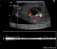
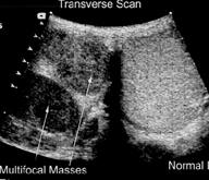
A testicular ultrasound (sonogram) is a test that uses reflected sound waves to produce a picture of the testicles and scrotum. An ultrasound can show the long, tightly coiled tube that lies behind each testicle and collects sperm (epididymis) and the tube (vas deferens) that connects the testicles to the prostate gland. The ultrasound does not use X-rays or other types of radiation.
A small handheld instrument called a transducer is passed back and forth over the scrotum. The transducer sends the sound waves to the computer which converts them into a picture that is displayed on a video monitor. The picture produced by ultrasound is called a sonogram, echogram, or scan. Pictures or videos of the ultrasound images may be saved as a permanent record.

Why It Is Done
Testicular ultrasound is done to:
- Evaluate a mass or pain in the testicles.
- Identify and monitor infection or inflammation of the testicles or epididymis.
- Identify twisting of the spermatic cord cutting off blood supply to the testicles (testicular torsion).
- Monitor for recurrence of testicular cancer.
- Locate an undescended testicle.
- Identify fluid in the scrotum (hydrocele), fluid in the epididymis (spermatocele), blood in the scrotum (hematocele), or pus in the scrotum (pyocele).
- Guide a biopsy needle for testicular biopsy when testing for infertility.
- Evaluate an injury to the genital area.
