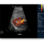
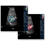
Renal Ultrasound

Acute flank pain and abdominal pain with hematuria are relatively common presenting complaints in the ultrasound department. Although urinary obstruction is a likely diagnosis in such patients, the differential diagnosis includes life-threatening disease processes, most importantly an expanding or ruptured abdominal aortic aneurysm. Emergency bedside sonography is a tool that can rapidly confirm the diagnosis of acute urinary obstruction and help exclude life-threatening processes.
It is important to know common medical terms used to describe the pathophysiology of urinary retention. The structural impediment to the flow of urine is termed obstructive uropathy. Unless this obstruction develops slowly it is typically painful, which is called renal colic. The most common cause is a kidney stone dislodged into the ureter called ureterolithiasis. Urine flow is blocked by the stone leading to back-up and dilation of the proximal ureter (hydroureter). As the obstruction progresses, more proximal structures like the renal collecting system (renal pelvis and calyces) becomes dilated, termed hydronephrosis. If the hydronephrosis is severe, the renal parenchyma becomes compressed and if lasting long enough (about 2-4 weeks), can cause loss of renal function.

As described above, the most common cause of renal colic and hydronephrosis is ureterolithiasis. But in general everything able to obstruct the inner lumen of the collecting system or causing extrinsic compression can block urinary flow and lead to renal colic.
Bedside renal sonography in the ultrasound department is also useful in the patient presenting with decreased urinary output or anuria, acute renal failure or pyelonephritis. Similar to the renal colic patient it allows the examiner to narrow the differential diagnosis by evaluating the retroperitoneal anatomical structures for abnormalities but gives only limited clues for the functional status of the urinary system.
